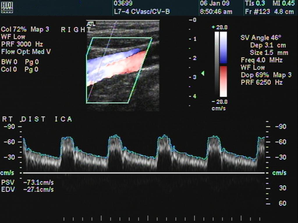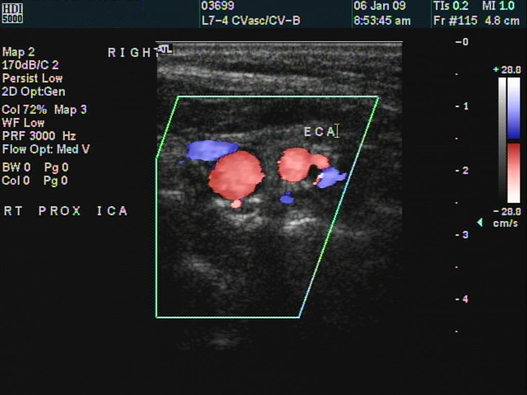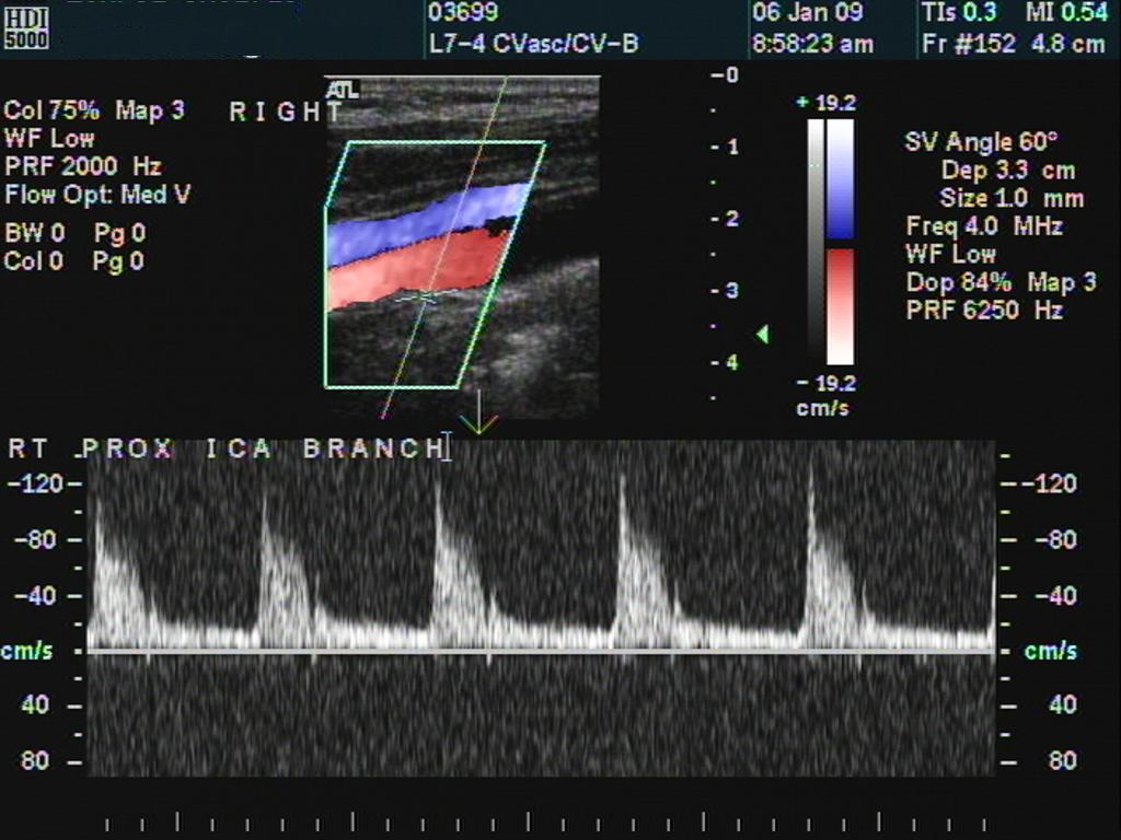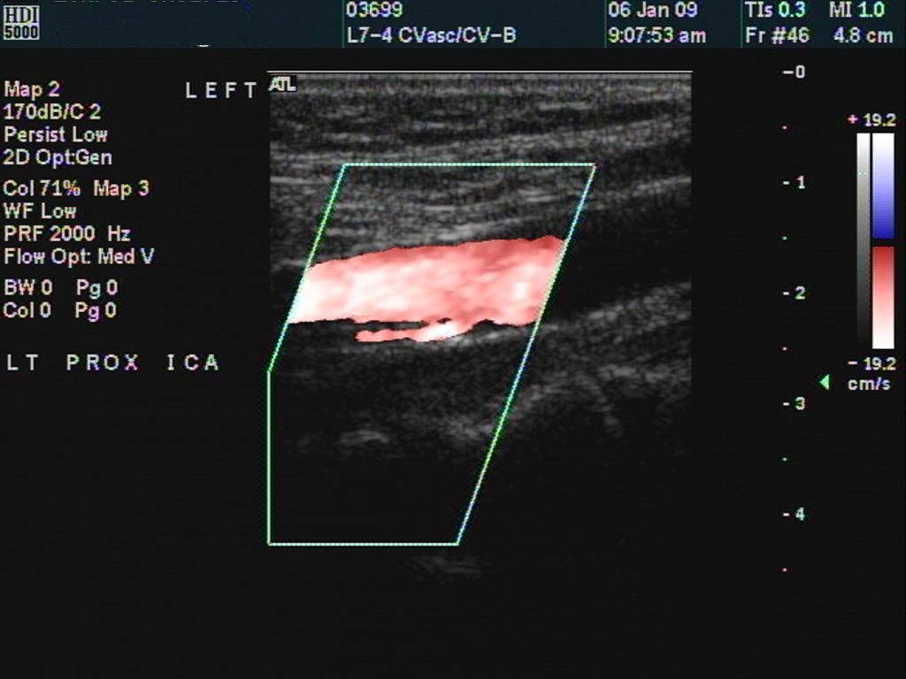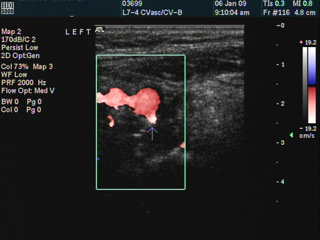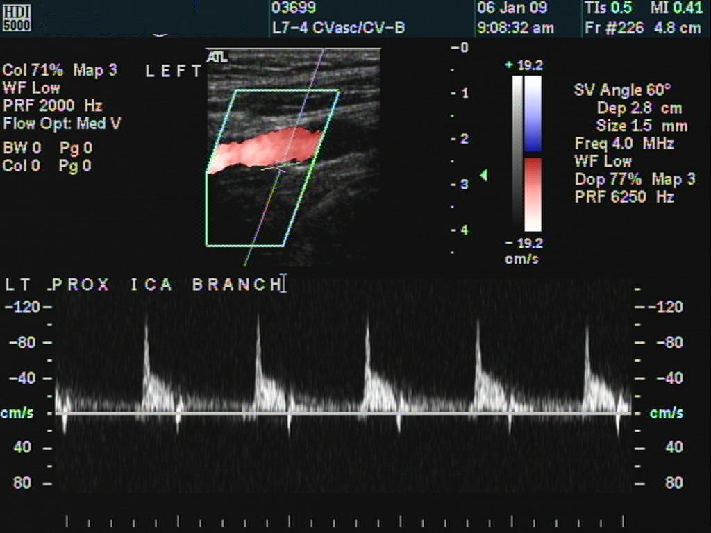

Case of the Month! Patient is a 67 y/o male that presented with a recent episode of diplopia lasting several minutes and then cleared. He had no recurrent episodes. On physical exam, he was also noted to a soft bilateral carotid bruit and so carotid duplex exam was ordered The technologists physical exam revealed - Radial pulses are equal; brachial blood pressure was 176/84 mm/Hg on the right and 180/84 mm/Hg on the left. A soft carotid bruit was appreciated bilaterally. Here are the findings - not a great diagnostic mystery but a very unusual finding! RIGHT BIFURCATION
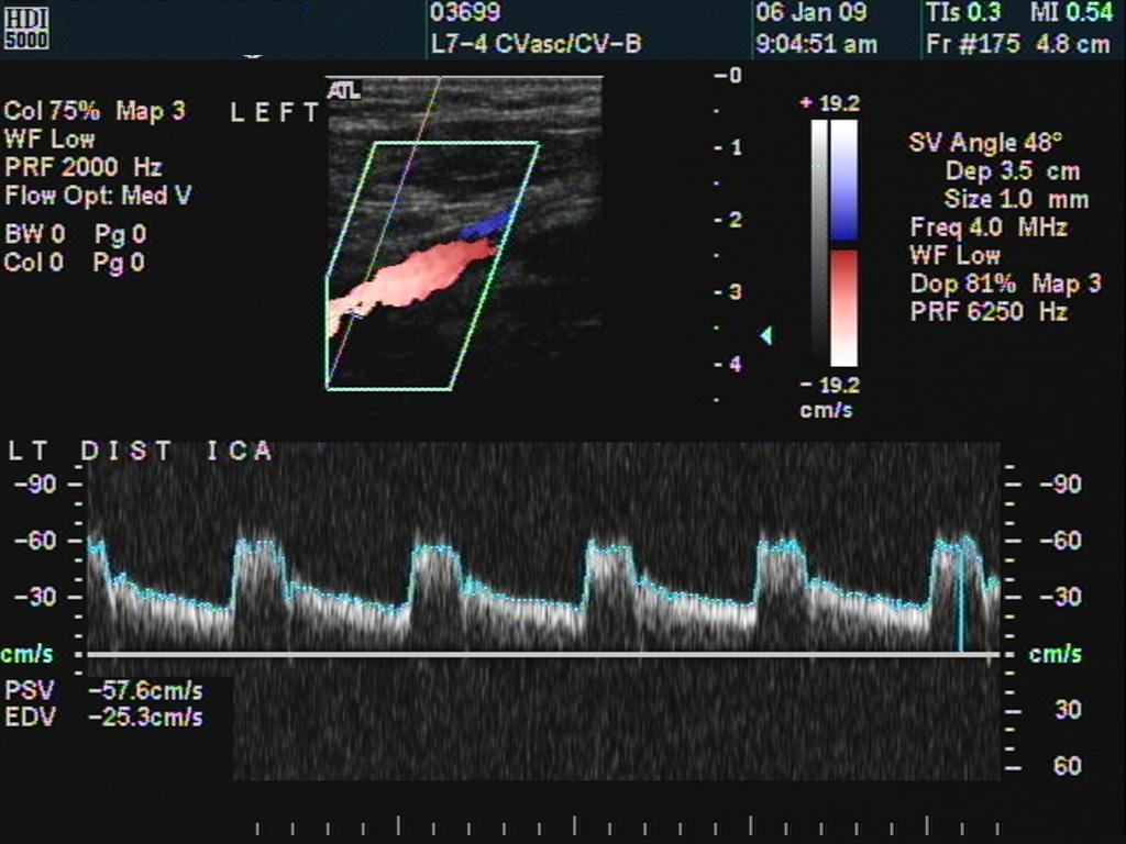
LEFT BIFURCATION
Discussion It is fairly clear that there is an internal carotid artery branch bilaterally in this otherwise completely normal study. This branch possibly represent an Anomalous ascending pharyngeal artery but this cannot really be proven with duplex. Anomalous branches of the internal carotid artery are quite rare and I was unable to find a "incidence" reported n the literature. Most cases reported in the literature involve branches in the intracranial circulation. In more than 25 years of carotid duplex scanning, this technologist has seen this anomaly three times (actually 4 as this would count as two!) One case more than 15 years ago involved a patient how has a short occlusion of the proximal internal carotid artery with continued patency distally via an anomalous proximal ICA branch. This patient underwent angiography which failed to show a patent ICA branch. Repeat duplex scanning confirmed its continued presence however as the patient was currently stable, he did not undergo endarterectomy. A similar case has been reported in the literature. http://download.journals.elsevierhealth.com/pdfs/journals/1533-3167/PIIS1533316708000228.pdf The presence of branches in the extracranial cerebrovascular vessels are a very important diagnostic finding. Virtually every anatomy text describes the ICA as having no extracranial branches, the first being the opthalmic artery which arises intracranially. Differentiation of the ICA from the external carotid artery is a common beginner mistake. Differentiation is typically easy in normal bifurcation (as in this case) but more difficult in diseased vessels. Useful tips:
I have typically used the presence of branches to be the best method for differentiation but in carotid duplex scanning, as in most things, there are few absolutes! I would love to hear of any cases |
|
