
Case of the Month!
Patient is a 66 y/o male who presented with a 1 week history of left lower extremity pain with multiple palpable lumps along the inside and back of his left calf. The palpable lumps were hard and tender and warm to touch. He has a history of deep vein thrombosis, twice, in the left leg approximately 20 years ago. Patient also has multiple large varicose veins.
What do you think? Discussion This patient has acute superficial thrombophlebitis in the small saphenous vein, which runs along the back of the calf, as well as several varicose veins. In other words he has a blood clot in the superficial venous system. Thrombosis in the superficial veins causes an inflammatory reaction that accounts for the pain, redness, and warmth. Treatment generally consists of anti-inflammatory medication and warm compresses. Varicose veins are the most common reason for development of a blood clot in a superficial vein. The tortuous dilatation of a varicosity allows for stasis or stagnation of blood and the risk of thrombosis rises dramatically. Superficial thrombophlebitis may extend up and down the saphenous vein or may remain confined to a cluster of tributary varicosities away from the main saphenous vein. It frequently is observed in varicose veins surrounding venous stasis ulcers. Superficial venous thrombosis may also occur from other reasons:
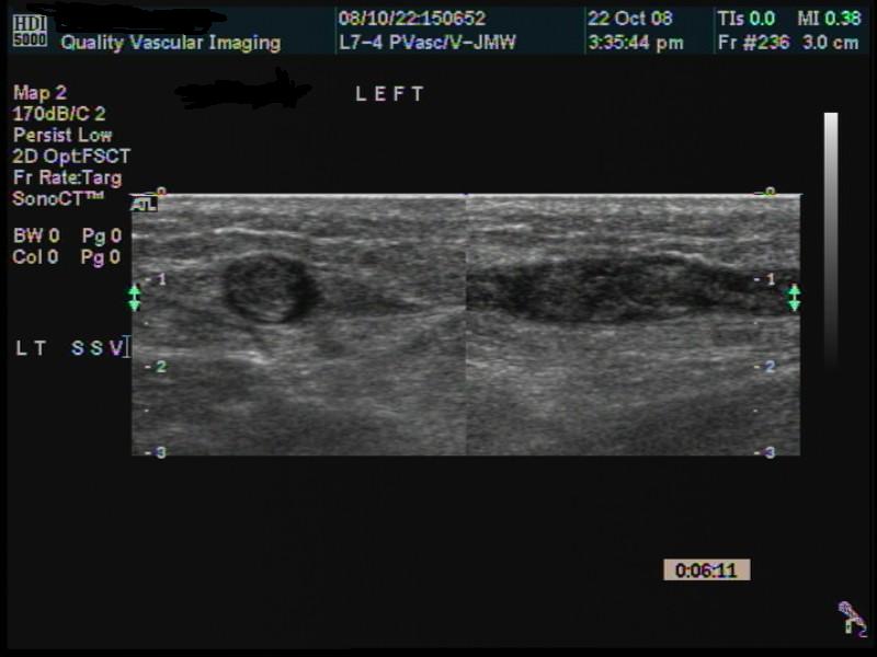
Diagnosis - In many cases, the diagnosis of superficial venous thrombosis can be made by a careful physical exam by an experienced clinician. Duplex ultrasound evaluation is the diagnostic study of choice for confirmation. Thrombosed veins may appear thickened on ultrasound, and the most consistent diagnostic finding is a lack of compressibility of the vein using the ultrasound transducer. An acutely thrombosed vein will often be dilated with low level echoes (hypoechoic) from within the lumen. An experienced vascular technologist should be able to diagnose superficial thrombophlebitis with very high sensitivity and specificity. Typically, the evaluation concerns the location and extent of superficial thrombosis, as well as the proximity to the deep venous system at the saphenofemoral or saphenopopliteal junction.
After an initial diagnosis of superficial thrombophlebitis, a duplex ultrasound examination may be be performed to look for progression of disease after treatment is initiated. A finding of no clot extension indicates successful therapy; thrombus extension or encroachment toward the deep venous system should prompt more aggressive treatment.
Treatment - The treatment of superficial venous thrombosis depends on its etiology, extent, and symptoms. Your physician can Duplex scanning gives an accurate appraisal of the extent of disease and thus allows determining more rational therapy. For the superficial, localized, mildly tender area of thrombophlebitis that occurs in a varicose vein, treatment with mild analgesics, such as aspirin, and the use of some type of elastic support are usually sufficient. Patients are typically encouraged to continue their usual daily activities. If extensive varicosities are present or if symptoms persist, phlebectomy or removal of the involved segment may be indicated. Please email me with any questions or comments. |
|


Compression Socks?
We carry the BEST!
Compression socks are not only a main stay in the treatment of venous and lymphatic disease but are now used by elite athletes everywhere. Please visit our webstore for more information about compression therapy by clicking the image above! .