
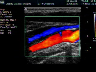
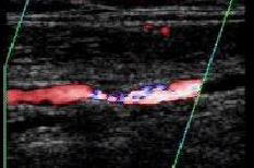
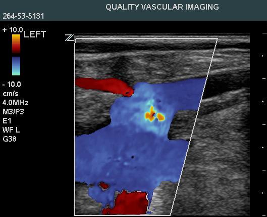
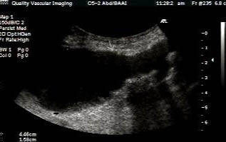
Duplex Ultrasound - is the primary testing instrument used in the evaluation of arteries and veins in a vascular laboratory. An ultrasound examination is a simple painless test and there is no special preparation unless we are evaluating abdominal vessels in which case you should fast after midnight. Ultrasound is a high frequency sound wave, similar to a sonar device. The sound waves bounce off the structures of interest, are reflected back, and are detected by the instrument. Two types of information are obtained, hence the term Duplex ultrasound - an image of the structures and information about blood flow. Different tissues reflect the ultrasound differently, so we can differentiate one from another. For example, the vessel wall appears different from the blood, the surrounding tissues, or plaque buildup inside the artery. At the same time, a special processing of the returning sound waves called Doppler allows us to get information about blood flow. Doppler is a physics principal that deals with the change in the frequency of sound waves because of motion. We know the frequency of the sound waves we send into the body. If they bounce off something that is moving, the frequency of the returning sound waves is changed. We compare the frequency of the sound waves we sent with the frequency of the returning sound waves and if different, we know they bounced off something moving, usually blood cells. If the blood flow is moving away from the transducer, the frequency is lower - if towards, the returning frequency is higher. In practice, we obtain an image of the blood vessel, surrounding tissues and any plaque or other abnormality inside along with simultaneous information about the blood flow. So if there is any plaque inside the artery, we can measure how much and importantly determine what effect it has on blood flow. In practice, a small plastic transducer will be moved along the area of interest. A gel is used to create good contact with the skin. Sometimes, the technologist may have to push slightly, you will not feel anything from the ultrasound, and you should experience no real discomfort. After you are gone, we perform a large number of additional measurements and calculations on the information we obtain and organize it into a report. The results will be reviewed by a physician and the report sent to your doctor. While your doctor will tell you what the findings mean to you, the technologist can provide much information about the disease and the testing process. You should feel free to ask questions. For a look at the image we see, click the video! You can click on any of the titles for detailed information! Carotid artery disease is the most common cause for stroke in the USA and early detection with accurate assessment is the key to prevention. At QVI, we have the most sophisticated equipment available for detecting this common condition. Our ultrasound instrument uses a super computer to compound multiple images that provides a unique view of plaque characteristics and surface irregularities. This allows an assessment of the risk a specific plaque presents to the patient. We are also able to evaluate the intracranial vessels for stenosis, arteriovenous malformation or vasospasm of the vessels within the skull. Traditionally, this information has only been obtainable by contrast angiography. Symptomatic patients without other explanation may benefit from this noninvasive test. Peripheral
Arterial Evaluation
Peripheral vascular disease in the legs presents as exertional leg pain known as claudication. This common condition can be suspected with a careful history and physical examination, but noninvasive testing can unequivocally establish its presence and quantitate the severity. In order to minimize costs, at QVI we start with the most inexpensive test that measures pressure and volume in various segments of the leg. Many times exercise is performed to elicit symptoms. If this is normal, often no further testing is needed. If abnormal, this can establish arterial insufficiency as the cause of the patients symptoms. If disease is present, all major arteries can be visualized using the color duplex scanner. The result is a detailed overview of the lower extremity arterial circulation. Venous duplex scanning is commonly performed on patients who present with a swollen leg to rule out the presence of deep vein thrombosis ("DVT"). Duplex scanning is the accepted “gold standard” for diagnosis of DVT. However, it is extremely operator dependent and the accuracy is primarily dependent upon the skill of the technologist performing the exam. There are a multitude of causes for leg swelling, for example venous insufficiency or synovial cysts. We are nationally recognized experts in venous testing with students coming from around the country for training at our facility. It is our philosophy that we provide an explanation for the patient’s symptoms, not just rule out a blood clot. You can be assured our patients will be fully evaluated, not just quickly scanned to “rule out DVT.” For additional information of veins, blood clots, and varicose veins. please visit the section on Venous evaluation.
Abdominal Aorta Evaluation
Aorta / iliac arteries - These are commonly evaluated with duplex scan for both blockage and aneurysm. While flow limiting stenosis of the abdominal aorta is quite rare, it is actually a very common area for development of atherosclerotic disease and the presence of disease here may important to know. Aneurysms is the primary reasons for testing and if identified, are closely followed for expansion.
|
QVI Home Virtual Vein Center Why QVI really is the Best in Vascular Ultrasound! Case of the Month Patients Referring Physicians Health Professionals Site Map |
Compression Socks?
We carry the BEST!
Compression socks are not only a main stay in the treatment of venous and lymphatic disease but are now used by elite athletes everywhere. Please visit our webstore for more information about compression therapy by clicking the image above! .