Venous Disease is a very common problem and can range from a minor annoyance to life threatening severity. There are two primary types of vein problems, venous insufficiency and venous thrombosis. Risk Factors - Venous disease affects people of all ages. There are significant risk factors for development of venous thrombosis and /or venous insufficiency.
Many of these can not be controlled, however by changing your lifestyle you can reduce the tendency to develop venous disease.
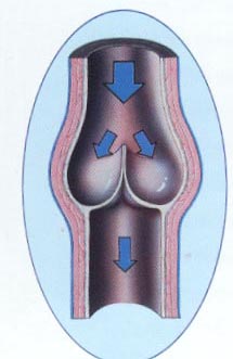
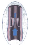
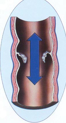
How the Veins Work - The veins of the body carry blood from the organs and tissues back to the heart in contrast to the arteries which carry blood to the organs. There are three types of veins - superficial, deep, and perforating veins. The superficial veins, lie just under the skin and are generally small and numerous. Their function is to drain blood from the skin and subcutaneous tissues. There are two main superficial veins called the saphenous veins. The deep veins are large veins deep in the muscle and they function as the primary conduit to carry blood out of the limb. They drain the muscles and organs as well as the superficial veins. Perforator veins connect the deep and superficial veins. Veins are elastic and collapsible; blood flows with very low pressure and against gravity. There are two mechanisms that work together in order to accomplish this function, the calf muscle pump and valves. As the calf muscle contracts, it squeezes the veins and pumps the blood out, this is referred to as the calf muscle pump. Valves are thin bicuspid leaflet like structures that open one way to allow blood to flow towards the heart and quickly close preventing blood from flowing backward. If a valve is not functioning, the blood is permitted to flow backward allowing the pooling of blood and resulting in vein distension.
Normal closed valve Normal open valve Damaged incompetent valve
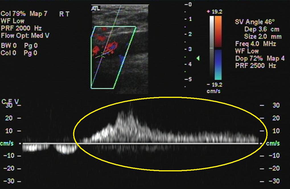
This is an ultrasound image of a femoral vein. The open valve is clearly visualized on the left and with inspiration, the valve closes on the right image. Venous insufficiency and valve function Veins are elastic and collapsible; blood flows with very low pressure and against gravity. There are two mechanisms that work together in order to accomplish this function, the calf muscle pump and valves . As the calf muscle contracts, it squeezes the veins and pumps the blood out, this is referred to as the calf muscle pump. Valves are thin bicuspid leaflet like structures that open one way to allow blood to flow towards the heart and quickly close preventing blood from flowing backward. If a valve is not functioning, the blood is permitted to flow backward allowing the pooling of blood and resulting in vein distension. Leaky one way valves cause what is called venous insufficiency. This can be thoroughly evaluated with duplex ultrasound - however this takes significant experience and skill. Most providers of vascular ultrasound services typically look for blood clots and unfortunately, many do a very marginal job evaluating venous insufficiency. QVI has extensive experience in evaluating this condition and provides training for technologists from around the country.
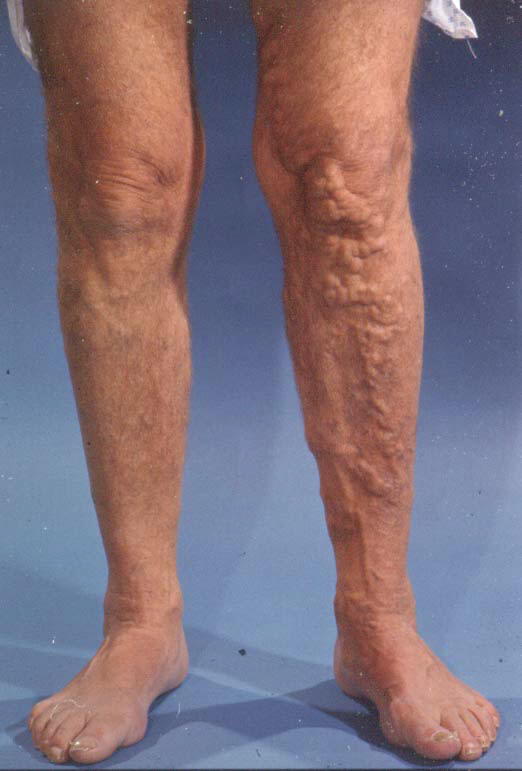
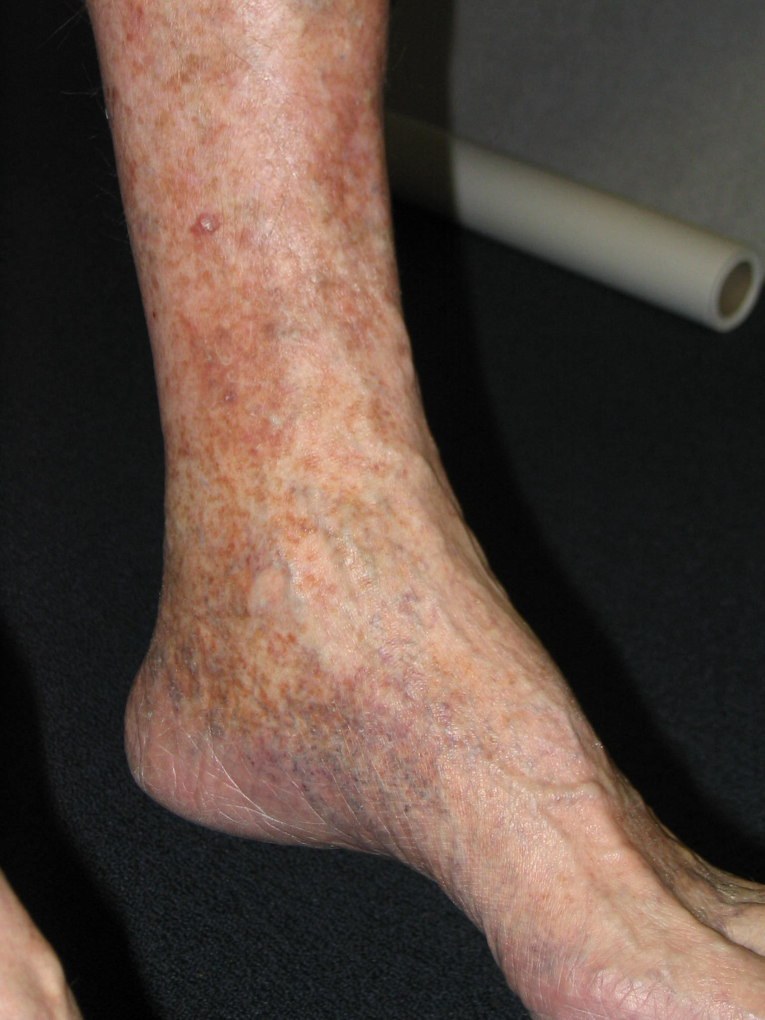
Superficial Veins - The superficial veins are poorly supported beneath the skin. When the valves begin to leak (venous insufficiency), an increased pressure is transmitted into the veins. Over time, the veins tend to enlarge and stretch. If the dysfunction occurs in the tiny superficial veins, spider veins form. There are several treatment options for spider veins including compression stockings, injection, and/or laser. What’s right for you depends upon many factors and should be discussed with your doctor. The larger superficial veins can be seen bulging and twisted beneath the skin (varicose veins). In addition to being unsightly, they can result in symptoms ranging from aching, a feeling of heaviness, tenderness, swelling in the in the limb to skin erosion and ulceration. As with spider veins, treatment options are varied and may include compression stockings, injection, laser, and a variety of surgical procedures depending upon many factors and the severity of the varicosities. See venous treatments In the case of ulceration, treatment is generally more aggressive. If insufficiency occurs in both the superficial and deep system compressive stockings are usually recommended.
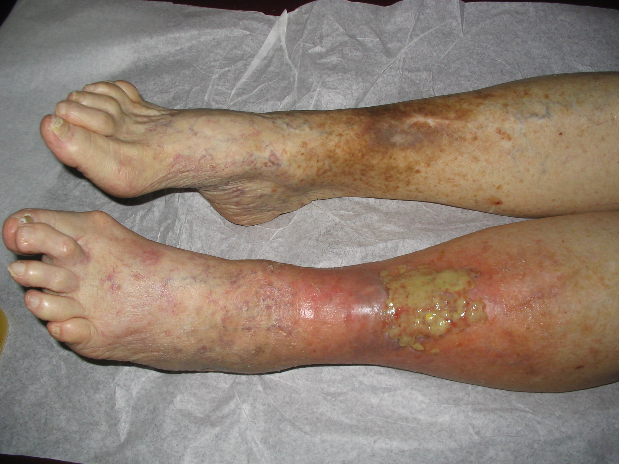
Venous thrombosis, or a blood clot in the vein, is an acute problem and as such, it is important treatment is sought quickly. If a blood clot is in the superficial veins, serious complications are rare, and treatment is conservative, often with anti-inflammatory medications. There may be localized pain or tenderness, swelling, and redness along the vein and a hard lump or cord may be felt under the skin. Deep veins Venous thrombosis in a deep vein is a very serious problem. Left untreated, there is a tendency for the clot to grow or propagate. The major potential danger is a blood clot breaking loose which lodges in the lungs preventing oxygen from entering the blood. This is called a pulmonary embolus and while it occurs in a relatively small percentage of patients with deep vein thrombosis (DVT), it can be fatal. Symptoms of DVT are variable depending upon the location and extent of the clot and whether the vein is partially or completely obstructed. This obstruction backs up blood behind the blockage as blood has to weave its way around the obstruction through other small veins. Swelling is a major symptom to suspect DVT. Pain and tenderness may also be present as a result of inflammation. Treatment of DVT usually consists of intravenous administration of a potent blood thinner to stop the clotting quickly. Many times this involves a hospital stay although some patients may be candidates for outpatient blood thinners. Once the blood is thinned to a “therapeutic level” an oral blood thinner is prescribed for an additional 3-6 months. The blood thinner prevents more clot from forming but does not generally dissolve the clot. The body can dissolve or lyse the clot over time. The formation of a blood clot results in a scarring of the walls of the vein and damage to valves. If extensive, this can result in what is called post phlebitic syndrome leading to discoloration of the skin and ultimately venous ulceration. Therefore, it is important to minimize the extent of the clot by timely treatment.
Compression
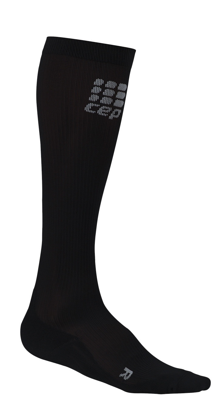
Stockings - Graduated compression stockings are often
recommended for a variety of venous conditions. We carry
Jobst and Medi graduated compression stockings! Compression
stockings create a gentle squeeze on the legs, preventing
venous distention and formation of varicose veins. They
come in a variety of styles and colors and are not what
most people typically think of when one mentions
compression stocking. If your physician recommends
compressive stockings, we can measure and fit you with the
best! See
compression stockings page!
For more information on purchasing quality
compression stockings, please visit:
www.compressionsocks.pro
| |||
QVI Home Virtual Vein Center Why QVI really is the Best in Vascular Ultrasound! Case of the Month Patients Referring Physicians Health Professionals Site Map |
Current Happenings

Introducing our new educational website.

Virtual Vein Center is a new concept in educational delivery. Get the education you need and want, when you need it. If you need CME, you can get them here as well.
To read more about it, click here for a complete page. Feel free to go to the site and browse around.


Several QVI staff took time to attend the 2014 American College of Phlebology Annual Congress in Phoenix Arizona in November to deliver numerous workshops and lectures. It was a high quality meeting as usual. The complete program is available for download here.


The 2014 SVU Annual Conference was held in Orlando and several QVI attended and presented numerous presentations. Jeannie was also honored as a Fellow of the SVU.
To read more

Jeannie recently attended the 25th Society of Vascular Medicine 2014 Annual Conference as an invited speaker in La Jolla, Ca. Her numerous lectures were very well received.

The International Union of Phlebology, in conjunction with the American College of Phlebology held its World Meeting in Boston in September 2013. Held only every 4 years, this was the first time ever in the US. Several QVI staff were invited speakers presenting some original scientific research.

Sydney, Australia

Bill was the International Keynote Speaker at the Australian Sonographer Association Annual National Conference in Sydney.
What a great experience!
To read more about this and our other international teaching


QVI was once again awarded the D.E. Strandness Award for Scientific Excellence at the 2013 SVU Annual Conference.
To read more -

Medical Compression socks continue to be on the forefront of venous treatment. Recently, they have entered the realm of the athlete. To learn more about what compression socks can do you you, please visit compressionsocks.pro


QVI wins the D.E Strandness Award at the 2012 SVU Annual Conference!
Read more about it!

To go to the
CASE OF THE MONTH!
Click the QVI logo

