
Carotid Artery Disease Carotid artery disease is the most common cause of stroke in the USA and early detection with accurate assessment is the key to prevention. How does the cerebrovascular system work? - Blood leaves the heart via the aorta and into the common carotid arteries (CCA) on each side of the neck. About half way up the neck, this artery branches into the external carotid (ECA) that supplies the face and scalp, and the internal carotid artery (ICA) that supplies the brain and the eye. The split or bifurcation is a common area for disease to occur. The vertebral arteries branch off the subclavian arteries, course along the spinal cord and supply the back of the brain. These 4 vessels make up the primary blood supply to the brain and these can be easily evaluated by noninvasive methods.
Atherosclerosis , commonly known as hardening of the arteries, is responsible for the majority of problems in the carotid arteries. Normally the inner wall of an artery called the intima is smooth and elastic, allowing blood to flow freely. In vessels affected by atherosclerosis, the intima becomes thickened and rough by a build up cholesterol or fatty materials. This build up, much like rust in a pipe, is called plaque. As this plaque increases, it obstructs the opening or lumen of the blood vessel and may alter or limit the flow of blood. If the artery is severely narrowed, the amount of blood getting past can be so limited, that various symptoms can result. However, as long as this is a gradual process, the body is often very good at developing alternative pathways for blood flow called collaterals. As the artery narrows, the same amount of blood wants to get through so it has to speed up, much like a “kink” in a garden hose. This fast, turbulent blood flow can break down the surface of the plaque and cause pieces of plaque called emboli to break loose. These can travel to the brain or the eye often with dire consequences. Turbulent blood flow can produce a sound called a bruit that can be heard with a stethoscope and is often the first sign of disease. Diagrams of increasing build-up of deposits within the artery wall, gradually bulging the inner layer of the artery wall into the lumen or opening inside the vessel resulting in a restriction of the blood flow. Small plaques that do not result in flow disturbances are unlikely to cause symptoms. In contrast, narrowings that limit the blood flow cause increased blood velocities and turbulent flow (like a kink in a garden hose.) This results in stresses on the inside layer of the vessel. If this lining breaks down, particles called emboli are carried to the brain and can result in a stroke. Symptoms -
Diagnosis - Diagnosis is most often made with ultrasound, a relatively inexpensive, completely safe and non-invasive technique that obtains pictures of the vessels and information about the blood flow. Plaque characteristics such as surface irregularities coupled with information about the blood flow allow an assessment of the risk a specific plaque presents to the patient. We are also able to evaluate the intracranial vessels for stenosis, arteriovenous malformation or vasospasm of the vessels within the skull. Traditionally, this information has only been obtainable by contrast angiography. Symptomatic patients without other explanation may benefit from this noninvasive test.
Figure 1 An ultrasound of the carotid artery bifurcation or branching showing widely patent arteries. However note the mild degree of wall thickening in this exceptional image. Figure 2 The internal carotid artery, which is the main blood supply to the brain, is shown with gray scale and color Doppler. The red demonstrates blood flow to the brain while the blue is flow in the jugular vein. Figure 3 Figure 4 Figure 3 - The internal carotid artery showing a large plaque along the back wall of the artery Figure 4 - This image of a carotid bifurcation uses a different Doppler processing the demonstrates the opening of the vessel extremely well. Note the "divot" which may represent an ulcer in the plaque. Figure 5 Figure 6 The above images show a color Doppler image of an internal carotid artery with a severe stenosis. Figure 6 shows the spectral analysis which is a detailed analysis of the blood flow showing a high velocity flow pattern due to the narroing in the artery. Treatments - Most patients with carotid artery disease do not have symptoms and are treated very conservatively without any intervention. But what else can be done??
Diagram of a Guidant "Acculink" carotid stent with a device to catch any pieces that could break loose during the procedure. |
QVI Home Virtual Vein Center Why QVI really is the Best in Vascular Ultrasound! Case of the Month Patients Referring Physicians Health Professionals Site Map |
Current Happenings

Introducing our new educational website.

Virtual Vein Center is a new concept in educational delivery. Get the education you need and want, when you need it. If you need CME, you can get them here as well.
To read more about it, click here for a complete page. Feel free to go to the site and browse around.


Several QVI staff took time to attend the 2014 American College of Phlebology Annual Congress in Phoenix Arizona in November to deliver numerous workshops and lectures. It was a high quality meeting as usual. The complete program is available for download here.


The 2014 SVU Annual Conference was held in Orlando and several QVI attended and presented numerous presentations. Jeannie was also honored as a Fellow of the SVU.
To read more

Jeannie recently attended the 25th Society of Vascular Medicine 2014 Annual Conference as an invited speaker in La Jolla, Ca. Her numerous lectures were very well received.


The International Union of Phlebology, in conjunction with the American College of Phlebology held its World Meeting in Boston in September 2013. Held only every 4 years, this was the first time ever in the US. Several QVI staff were invited speakers presenting some original scientific research.

Sydney, Australia

Bill was the International Keynote Speaker at the Australian Sonographer Association Annual National Conference in Sydney.
What a great experience!
To read more about this and our other international teaching


QVI was once again awarded the D.E. Strandness Award for Scientific Excellence at the 2013 SVU Annual Conference.
To read more -

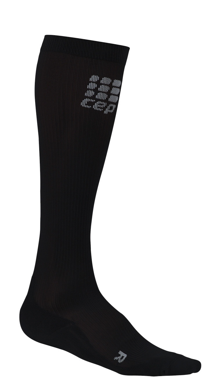
Medical Compression socks continue to be on the forefront of venous treatment. Recently, they have entered the realm of the athlete. To learn more about what compression socks can do you you, please visit compressionsocks.pro


QVI wins the D.E Strandness Award at the 2012 SVU Annual Conference!
Read more about it!

To go to the
CASE OF THE MONTH!
Click the QVI logo


