
QVI Case of the Month! For past cases,
click here A 68 y/o male presents with right thigh swelling and bruising due to recent cardiac catheterization via the right femoral artery. Due to these symptoms, he had already undergone two previous ultrasound examinations performed elsewhere at two different facilities, both of which were reported to be within normal limits. He has a history of atrial fibrillation, hypertension, hyperlipidemia, and a remote history of tobacco abuse.
The technologist clinical exam revealed the following: The right thigh was extensively bruised from supra-inguinal region to the distal thigh. Significant swelling of the right thigh was also noted.
Review the following images and find the results below:
Figure 1 Figure 2
Figure 3 Figure 4
Figure 5 Figure 6
Operative Findings: The patient was explored surgically. The mass in the right groin was found to be a mass of matted inflamed lymphnodes; therefore, a superficial femoral lymphadenectomy was performed. A right superficial femoral artery to common femoral vein arterial venous fistula (AVF) was found. A right common femoral deep venous thrombosis was also identified. An angioseal was inserted in the common femoral artery, and a greenfield filter was inserted in the inferior vena cava (IVC).
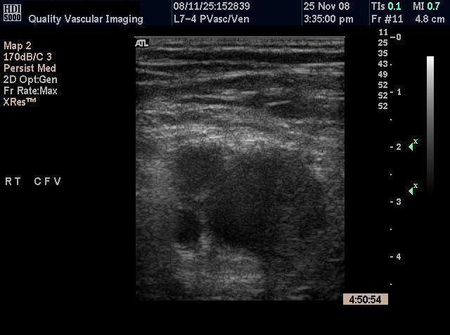
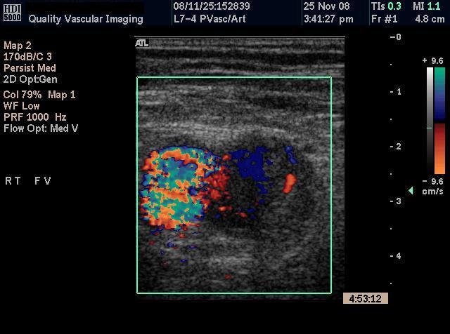
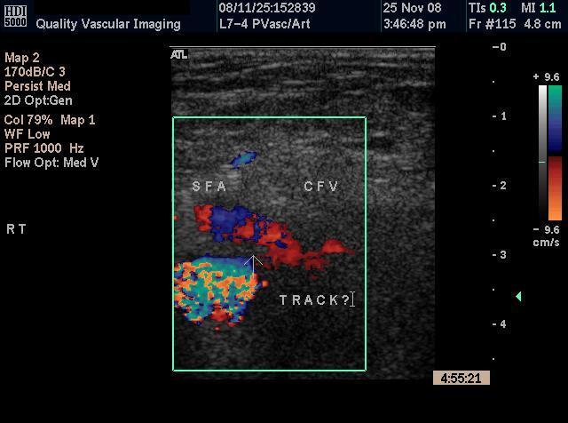
Discussion: The US findings show a greatly dilated “mass” medial to the superficial femoral and profunda femoris arteries (Figs. 1 & 2) and could be either a large mass / pseudoaneurysm or the common femoral vein. At first glance, this could be mistaken as a pseudoaneurysm (or “false aneurysm”) so named from the finding of a pulsatile mass on physical exam. A pseudoaneurysm is the result of trauma to all three layers of an artery resulting in a hematoma that contains the bleed. The hematoma must continue to communicate with the artery to be considered a pseudoaneurysm. A pseudoaneurysm differs from a true aneurysm in that a pseudoaneurysm does not contain any of the vessel wall. Femoral pseudoaneurysms may complicate up to 8% of vascular interventional procedures. Small pseudoaneurysms can spontaneously clot, while others need definitive treatment. Many institutions now close these with thrombin injection under ultrasound guidance.
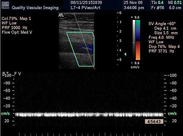
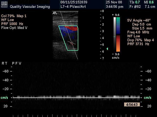
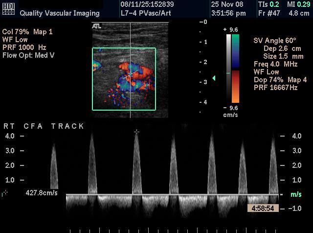
Generally we know of pseudoaneurysms to have a classic “to and fro” spectral waveform from within the track from the artery to the sac and interestingly, this classical waveform is clearly seen above; however, direct visualization of a track between the femoral artery and the common femoral vein in transverse is identified with color Doppler. Spectral analysis obtained in the track revealed greatly elevated peak systolic velocities of greater than 400 cm/sec with a prominent reversed diastolic component quite similar to what would be expected in a pseudoaneurysm. In fact, this is an atypical presentation of an arteriovenous fistula. An arteriovenous (AV) fistula is defined as an abnormal communication between the arterial and venous system. It may be congenital, or acquired secondary to trauma, tumors, or as a result of surgery. Although the physiological effects of traumatic fistulas have been well characterized, their clinical manifestations are quite variable due to differences in location, size, and duration. Duplex findings in a typical, traumatic AVF would generally have a continuous and pulsatile, high velocity flow. Additionally, upstream venous flow will often be pulsatile. When looking at the common femoral vein in transverse (Fig1) it clearly appears distended and spectral analysis demonstrates continuous flow throughout this segment. (Fig3) It could be surmised that the distention of the vein may be caused but high arterial pressure transmitted to the vein from the superficial femoral artery and the continuous flow may be caused by extrinsisc compression of the superficial femoral mass. However, this was confirmed at surgery to be a dep venous thrombosis. When looking at the transverese gray scale image, one can see thrombus within the lumen. Generally, we sue spectral analysis in the venous system to help confirm an AVF. In this case, we surmise that the limited outflow due to the deep venous obstruction as well as possible extrinsisc compression resulted in a low velocity continuous flow. Overall, a very atypical presentation for a traumatic AVF!
For past cases,
click here |
QVI Home Virtual Vein Center Why QVI really is the Best in Vascular Ultrasound! Case of the Month Patients Referring Physicians Health Professionals Site Map |

Current Happenings

Introducing our new educational website.
![]()
Virtual Vein Center is a new concept in educational delivery. Get the education you need and want, when you need it. If you need CME, you can get them here as well.
To read more about it, click here for a complete page. Feel free to go to the site and browse around.


Several QVI staff took time to attend the 2014 American College of Phlebology Annual Congress in Phoenix Arizona in November to deliver numerous workshops and lectures. It was a high quality meeting as usual. The complete program is available for download here.
The 2014 SVU Annual Conference was held in Orlando and several QVI attended and presented numerous presentations. Jeannie was also honored as a Fellow of the SVU.
To read more
Jeannie recently attended the 25th Society of Vascular Medicine 2014 Annual Conference as an invited speaker in La Jolla, Ca. Her numerous lectures were very well received.
The International Union of Phlebology, in conjunction with the American College of Phlebology held its World Meeting in Boston in September 2013. Held only every 4 years, this was the first time ever in the US. Several QVI staff were invited speakers presenting some original scientific research.
Sydney, Australia
Bill was the International Keynote Speaker at the Australian Sonographer Association Annual National Conference in Sydney.
What a great experience!
To read more about this and our other international teaching
QVI was once again awarded the D.E. Strandness Award for Scientific Excellence at the 2013 SVU Annual Conference.
To read more -
Medical Compression socks continue to be on the forefront of venous treatment. Recently, they have entered the realm of the athlete. To learn more about what compression socks can do you you, please visit compressionsocks.pro

QVI wins the D.E Strandness Award at the 2012 SVU Annual Conference!
Read more about it!
To go to the
CASE OF THE MONTH!
Click the QVI logo
Compression Socks?
We carry the BEST!
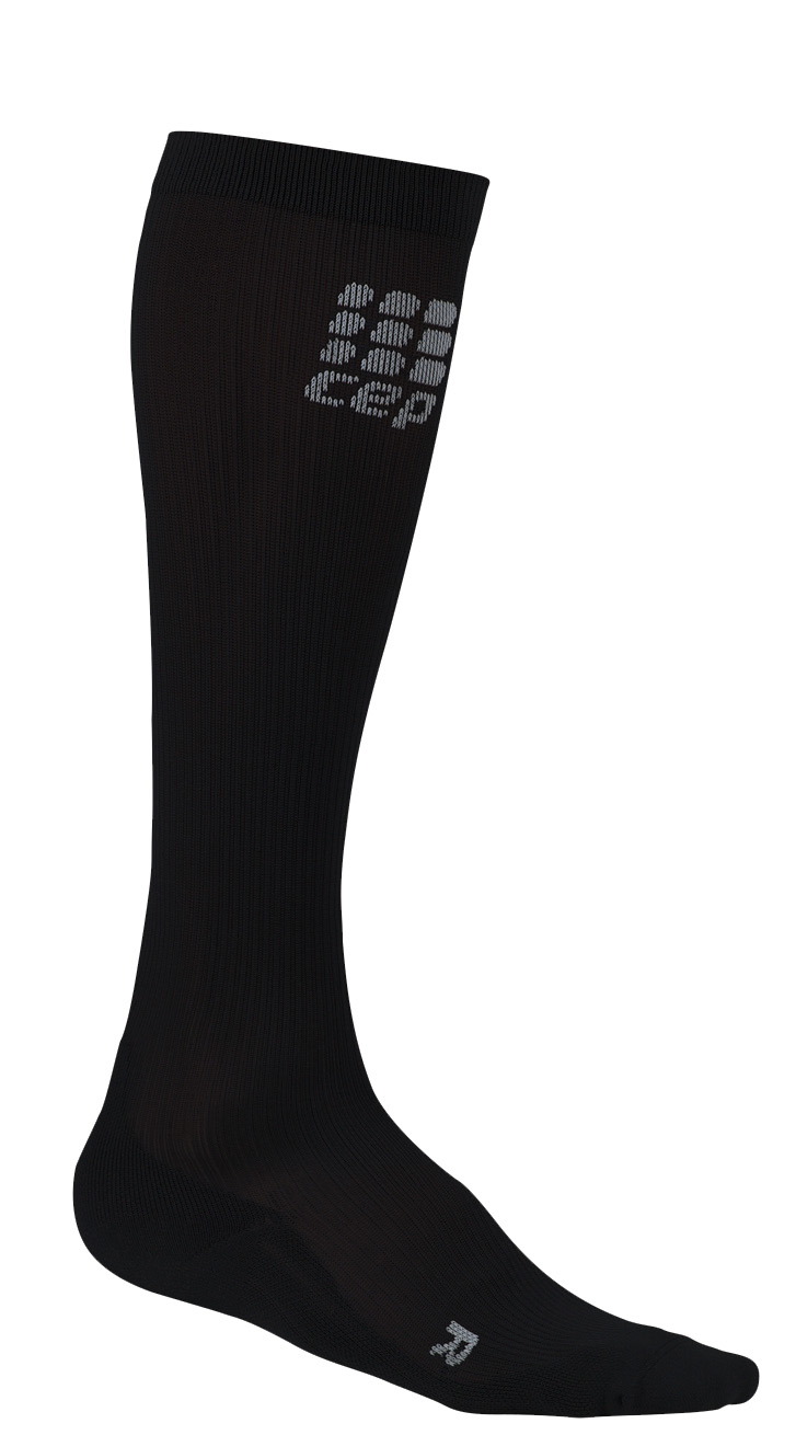
Compression socks are not only a main stay in the treatment of venous and lymphatic disease but are now used by elite athletes everywhere. Please visit our webstore for more information about compression therapy by clicking the image above! .